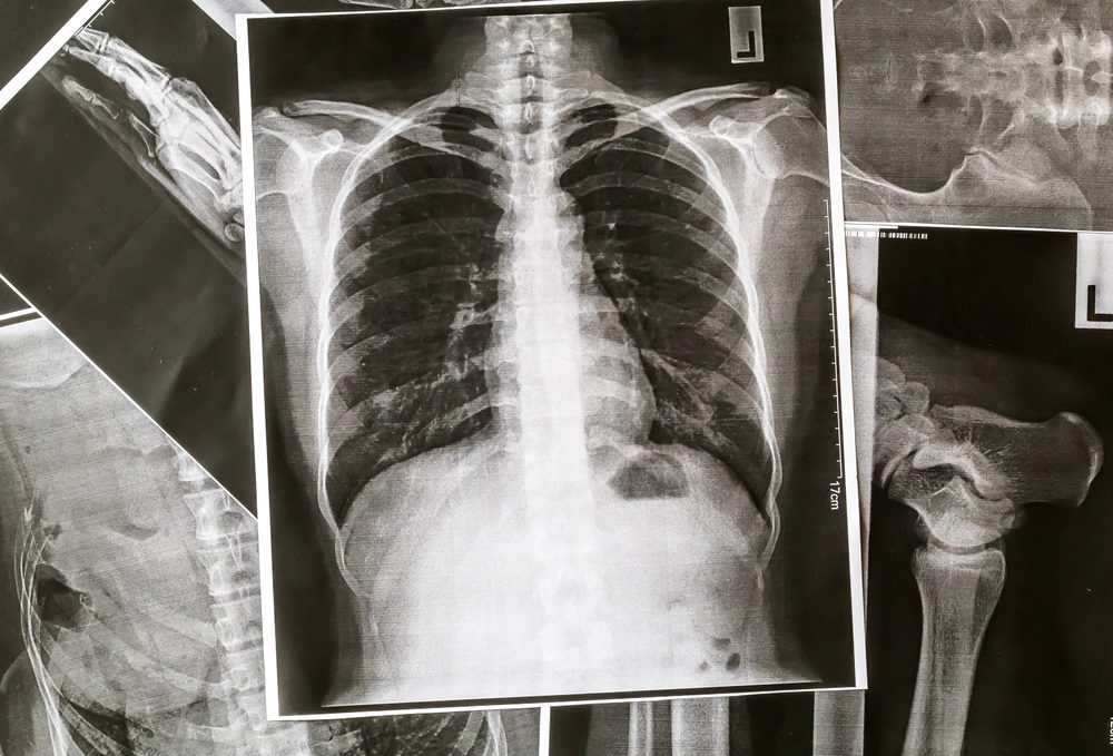
Spot in Lung on Chest X-ray Common and Typically Noncancerous
In fact, so many people have spots on their lungs that they show up on about 50 percent of all chest CT scans and 0.2% percent of chest x-rays. In fact, some estimate that more than half (PDF link) of all adults over the age of 50 have nodules on their lungs. Patients learning they have a 'spot' on their lung often assume they have cancer, according to a new study - but that's usually not the case. Researchers say doctors could do more to help patients. Cancer Research UK is a registered charity in England and Wales (1089464), Scotland (SC041666), the Isle of Man (1103) and Jersey (247). A company limited by guarantee. Registered company in England and Wales (4325234) and the Isle of Man (5713F). Registered address: 2 Redman Place, London, E20 1JQ. A lung nodule is defined as a 'spot' on the lung that is 3 centimeters (about 1.5 inches) in diameter or less. These nodules are often referred to as ' coin lesions ' when described on an imaging test.
December 30, 2011
Dear Mayo Clinic:
A spot in my lung showed up on a routine chest X-ray. I assumed it would be cancer, but my doctor says it may be something else. What else could it be?
Answer:
A solitary spot on a chest X-ray — generally referred to as a lung nodule — sometimes can be an early cancer, so it's important to talk to your doctor to determine the best course of action.
Lung nodules are common and typically noncancerous (benign). Among the most common causes of noncancerous lung nodules are scars or marks from a prior fungal infection, such as histoplasmosis or coccidiodomycosis, a bacterial infection, a mycobacterial infection such as tuberculosis, or a benign tumor called a hamartoma.
As in your situation, solitary lung nodules often are detected on an X-ray done for another reason, and appear as round, white shadows on an X-ray or computerized tomography (CT) scan. Lung nodules are common. If you have a CT scan of the chest, about half the time, one or more small nodules will be found. They appear white because they are denser than the surrounding lung, which is full of air and appears dark.
Generally, the smaller the nodule the less likely it may be cancerous (malignant). Nodules that measure less than 5 millimeters (mm) — or about 1/5 of an inch — are extremely likely to be benign. Those measuring more than 20 mm, which is about 3/4 of an inch, have a greater than 50 percent likelihood of being cancerous.
Along with size, several additional factors are considered when a solitary lung nodule is discovered.
For example, being 50 or older increases the probability that the nodule is cancerous. A history of smoking increases lung cancer risk. Asbestos exposure and a family history of lung cancer also are risk factors. Your doctor may compare your current X-ray or CT scan with a previous one to see if the nodule was present. If it was and there's no change in size, shape or appearance, chances are high it's noncancerous. Periodic imaging tests such as CT or other scans may be all that's needed to monitor for any changes. If there's no change after two years, follow-up is usually stopped.
Further testing may be recommended if the nodule is new or has changed in size, shape or appearance. Tests may include the following:
- CT scan of the chest if the nodule was first seen on a chest X-ray;
- Positron emission tomography (PET) scan to see how active the nodule cells are — high cellular activity is an indicator of cancer or active inflammation; and/or
- Tissue sample (biopsy the nodule).
A nodule that grows over time is more likely to be malignant, though some benign nodules can grow. When a nodule is a lung cancer that hasn't spread, it's at a stage that can usually be cured with surgery.

— David Midthun, M.D., Pulmonary and Critical Care Medicine, Mayo Clinic, Rochester, Minn.
Medically reviewed by Drugs.com. Last updated on Feb 5, 2020.

— David Midthun, M.D., Pulmonary and Critical Care Medicine, Mayo Clinic, Rochester, Minn.
Medically reviewed by Drugs.com. Last updated on Feb 5, 2020.
- Health Guide
What is Adenocarcinoma of the lung?
Adenocarcinoma of the lung is a type of non-small cell lung cancer. Casino 1995 watch online. It occurs when abnormal lung cells multiply out of control and form a tumor. Eventually, tumor cells can spread (metastasize) to other parts of the body including the
- lymph nodes around and between the lungs
- liver
- bones
- adrenal glands
- brain.
Compared with other types of lung cancer, adenocarcinoma is more likely to be contained in one area. If it is truly localized, it may respond to treatment better than other lung cancers.
Adenocarcinoma is the most common form of lung cancer. It's generally found in smokers. However, it is the most common type of lung cancer in nonsmokers. It is also the most common form of lung cancer in women and people younger than 45.
As with other forms of lung cancer, your risk of adenocarcinoma increases if you
- Smoke. Smoking cigarettes is by far the leading risk factor for lung cancer. In fact, cigarette smokers are 13 times more likely to develop lung cancer than nonsmokers. Cigar and pipe smoking are almost as likely to cause lung cancer as cigarette smoking.
- Breathe tobacco smoke. Nonsmokers who inhale fumes from cigarette, cigar, and pipe smoking have an increased risk of lung cancer.
- Are exposed to radon gas. Radon is a colorless, odorless radioactive gas formed in the ground. It seeps into the lower floors of homes and other buildings and can contaminate drinking water. Radon exposure is the second leading cause of lung cancer. It's not clear whether elevated radon levels contribute to lung cancer in nonsmokers. But radon exposure does contribute to increased rates of lung cancer in smokers and in people who regularly breathe high amounts of the gas (miners, for example). You can test radon levels in your home with a radon testing kit.
- Are exposed to asbestos. Asbestos is a mineral used in insulation, fireproofing materials, floor and ceiling tiles, automobile brake linings, and other products. People exposed to asbestos on the job (miners, construction workers, shipyard workers, and some auto mechanics) have a higher-than-normal risk of lung cancer. People who live or work in buildings with asbestos-containing materials that are deteriorating also have an increased risk of lung cancer. In addition to having a higher risk of adenocarcinoma, people who have been exposed to asbestos have a greater risk of developing mesothelioma. This is a type of cancer that starts in the tissue surrounding the lungs. Mesothelioma can also arise in tissue that surrounds organs within the abdomen.
- Are exposed to other cancer-causing agents at work. These include uranium, arsenic, vinyl chloride, nickel chromates, coal products, mustard gas, chloromethyl ethers, gasoline, and diesel exhaust.
Symptoms
Many people with adenocarcinoma of the lung or other types of lung have no symptoms. It may be detected on chest x-ray or CT scan that is performed for screening or some other medical reason.
All lung cancers, including adenocarcinoma, have similar symptoms. They include
- a cough that doesn't go away
- coughing up blood or mucus
- wheezing
- shortness of breath
- trouble breathing
- chest pain
- fever
- discomfort when swallowing
- hoarseness
- weight loss
- poor appetite.
If the cancer has spread beyond the lungs, it can cause other symptoms. For example, you may have bone pain if it has spread to your bones.
Many of these symptoms can be caused by other conditions. See your doctor if you have symptoms so that the problem can be diagnosed and properly treated.
Diagnosis
2 Spots On Lungs
Your doctor will start by taking your health history. He or she will ask about your smoking habits and whether you live with a smoker. Your doctor also will ask whether you may have been exposed to asbestos or other cancer-causing agents at work.
Next, he or she will order imaging tests to check your lungs for masses. In most cases, a chest x-ray will be done first. If the x-ray shows anything suspicious, a CT scan will be done. As the scanner moves around you, it takes many pictures. A computer then combines the images. This creates a more detailed image of the lungs, allowing doctors to confirm the size and location of a mass or tumor.
You may also have a magnetic resonance imaging (MRI) scan or a positron emission tomography (PET) scan.
MRI scans provide detailed pictures of the body's organs, but they use radio waves and magnets to create the images, not x-rays.
PET scans look at the function of tissue rather than anatomy. Lung cancer and many other cancers show intense metabolic activity on a PET scan. Radioactive sugar is injected into a vein. Cancer cells are more active and need more sugar than surrounding tissue. This makes areas of cancer light up more brightly on the scan.
If cancer is suspected based on these images, more tests will be done to make the diagnosis, determine the type of cancer, and see if it has spread. These tests may include the following:
- Sputum sample — Coughed-up mucus is checked for cancer cells.
- Biopsy — A sample of abnormal lung tissue is removed and examined under a microscope in a laboratory. The tissue is often obtained during a bronchoscopy. However, surgery may be necessary to expose the suspicious area.
- Bronchoscopy — During this procedure, a tube-like instrument is passed down the throat and into the lungs. A camera on the end of the tube allows doctors to look for cancer and to remove a small piece of tissue for a biopsy.
- Mediastinoscopy — In this procedure, a tube-like instrument is used to biopsy lymph nodes or masses between the lungs. (This area is called the mediastinum.) A biopsy obtained this way can diagnose the type of lung cancer and determine whether the cancer has spread to lymph nodes.
- Fine-needle aspiration — With a CT scan, a suspicious area can be identified. A tiny needle is then inserted into that part of the lung. The needle removes a bit of tissue for examination in a laboratory. The type of cancer can then be diagnosed.
- Thoracentesis — If there is fluid build-up in the chest, it can be drained with a sterile needle. The fluid is then checked for cancer cells.
- VATS (video-assisted thoracoscopy) — In this procedure, a surgeon inserts a flexible tube with a video camera on the end into the chest through an incision. He or she can then look for cancer in the space between the lungs and the chest wall. Abnormal lung tissue can also be removed.
- CT, PET, and bone scans — These imaging tests can detect lung cancer that has spread to the brain, bones, or other parts of the body.
- Thoracotomy. On occasion, a larger incision in the chest may be required to get tissue for examination in the laboratory.
After the cancer has been diagnosed, it is assigned a 'stage.' The stage indicates the tumor's size and how far it has spread. Stages I through III are further divided into 'A' and 'B' categories. Stage I tumors are small and have not invaded surrounding tissues. Stage II and III tumors have invaded surrounding tissues and/or organs and have spread to lymph nodes. Stage IV tumors have spread beyond the chest.
Expected Duration
Adenocarcinoma of the lung will continue to grow and spread until it is treated.
Prevention
To reduce your risk of adenocarcinoma and other forms of lung cancer,
- Don't smoke. If you already smoke, talk to your doctor about getting the help you need to quit.
- Avoid secondhand smoke. Choose smoke-free restaurants and hotels. Ask guests to smoke outdoors, especially if there are children in your home.
- Reduce exposure to radon. Have your home checked for radon gas. A radon level above 4 picocuries/liter is unsafe. If you have a private well, have your drinking water checked, too. Kits to test for radon are widely available.
- Reduce exposure to asbestos. Because there is no safe level of asbestos exposure, any exposure is too much. If you have an older home, check to see if any insulation or other asbestos-containing material is exposed or deteriorating. The asbestos in these areas must be professionally removed or sealed up. If the removal isn't done properly, you may be exposed to more asbestos than you would have been if it has been left alone. People who work with asbestos-containing materials should use approved measures to limit their exposure and to prevent bringing asbestos dust home on their clothing.
- The U.S. Preventive Services Task Force recommends annual screening for lung cancer with low-dose computed tomography in adults ages 55 to 80 years:
- Have a 30 pack-year smoking history (pack years is calculated by multiplying the number of cigarettes smoked per day times the number of years you smoked), AND
- Are currently smoke or have quit within the past 15 years, AND
- Are healthy enough to undergo lung cancer surgery.
Treatment
Treatment depends on the cancer's stage as well as the patient's condition, lung function, and other factors. (Some patients may have other lung conditions, such as emphysema or COPD—chronic obstructive pulmonary disease.) If the cancer has not spread, surgery is usually the treatment of choice. There are three types of surgery:
- Wedge resection removes only a small part of the lung.
- Lobectomy removes one lobe of the lung.
- Pneumonectomy removes an entire lung.
Lymph nodes are also removed and examined to see if the cancer has spread.
Some surgeons use video-assisted thoracoscopy (VATS) to remove small, early-stage tumors, especially if the tumors are near the outer edge of the lung. (VATS can also be used to diagnose lung cancer.) Because the incisions for VATS are small, this technique is less invasive than a traditional 'open' procedure.
Because surgery will remove part or all of a lung, breathing may be more difficult afterwards, especially in patients with other lung conditions (emphysema, for example). Doctors can test lung function prior to surgery and predict how it might be affected by surgery.
Depending on how far the cancer has spread, treatment may include chemotherapy (the use of anticancer drugs) and radiation therapy. These may be given before and/or after surgery.
When the tumor has spread significantly, chemotherapy may be recommended to slow its growth, even if it cannot cure the disease. Chemotherapy has been shown to ease symptoms and prolong life in cases of advanced lung cancer.
Radiation therapy can relieve symptoms, too. It is often used to treat lung cancer that has spread to the brain or bones and is causing pain. It can also be used alone or with chemotherapy to treat the lung cancer that is confined to the chest.
People who may not withstand surgery due to other serious medical problems may receive radiation therapy, with or without chemotherapy, to shrink the tumor.
Today, cancerous tissue may be tested for specific genetic abnormalities (mutations). Doctors may then be able to treat the cancer with a 'targeted therapy.' These therapies can derail the cancer's growth by preventing or changing chemical reactions linked to particular mutations. For example, some target therapies prevent cancer cells from receiving chemical 'messages' telling them to grow.
Knowing about specific genetic mutations can help predict which therapy will be best. This strategy can be especially helpful in certain patients, such as women with adenocarcinoma of the lung who have never smoked. The study of these mutations is usually performed in patients with lung adenocarcinomas.
New discoveries regarding the different mutations continue to improve therapy for adenocarcinoma of the lung in the future. Initial therapy or subsequent therapy may not necessarily require the use of chemotherapy.
Even after treatment has been completed, lung cancer patients must return for regular follow-up appointments. Even if the cancer was initially placed into remission, it can return months or even years later.
When To Call a Professional
Call your doctor promptly if you have any symptoms of lung cancer, especially if you smoke or have been exposed to asbestos.
2 Inch Spot On Lung
Prognosis
The outlook depends on the cancer's stage, the ability to identify specific mutations of the cancer cells for targeted therapy, and the patient's overall health. Adenocarcinoma of the lung can be cured if the entire tumor is removed surgically or destroyed with radiation. Overall, the prognosis for lung cancer that has spread is still poor. However, newer therapies have helped certain subsets of patients extend their lives.
2 Spots Of Cancer On Lungs
External resources
National Cancer Institute (NCI)
https://www.nci.nih.gov/
American Cancer Society (ACS)
https://www.cancer.org/
2 Spots On Lungs
American Lung Association
https://www.lung.org/ Slots of vegas free spin codes.
National Heart, Lung, and Blood Institute (NHLBI)
https://www.nhlbi.nih.gov/
U.S. Environmental Protection Agency (EPA)
https://www.epa.gov/
Further information
2 Spots On Lung
Always consult your healthcare provider to ensure the information displayed on this page applies to your personal circumstances.
